
Unleashing the True Potential of Human
Brain Research
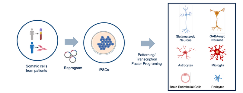
The basis of the NeuCyte technology is the proprietary methods for producing SynFire® cells. Derived from induced pluripotent stem cells (iPSCs), large homogenous batches of multiple neuronal subtypes with high functionality can be created.


Patient
Reprogram
iPS Cells
Differentiate
Pure Populations of Different Neural Subtypes
Defined iN/Astroglial Co-culture
Molecular Analyses
and Functional Studies
Phenotypic Screening and Drug Development
Neurotoxicity and Safety Assesment
New Drug
Cell-based Disease Modeling
Relevant In Vitro
Pre-Clinical Studies
SynFire iNs represent a versatile in vitro cell system for basic research and disease modeling, drug discovery and development. These cells are suitable for a variety of functional assays. For example, the effect of compounds on neuronal survival, axonal outgrowth, or dendritic arborization can be measured by standard assessment of viability, or image-based analysis of labeled cells, respectively. When co-cultured with glial cells, effects on synapse formation and composition, transcriptional programs, and electrophysiology can be tested.


tdTomato/TUJ1/ DAPI

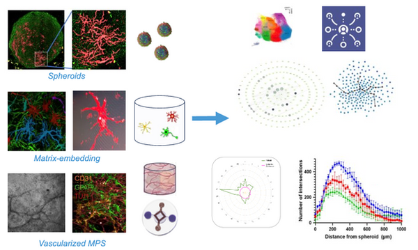
Based on the SynFire technology, complex human neural in vitro systems can be established to assess relevant higher order electrophysiology readouts, which allow for a more reliable predictor of drug efficacy and potential neural safety/toxicology. NeuCyte's unique human neural platform allows for the evaluation of human specific neural phenotypes that are not identifiable in traditional models.

SynFire excitatory and inhibitory neurons are combined with primary astrocytes within a three-dimensional brain assembloid, establishing a microphysiological system (MPS) platform that better mimics the complexity of the human brain with low assembloid to assembloid variance. As shown, neurons (TUJ1), astrocytes (GFAP) and cell nuclei (DAPI) can be visualized within assembloids by immunocytochemistry. Therefore, high content imaging of the SynFire platform can be utilized to identify morphological phenotypes associated with neurological diseases that are captured in a three-dimensional context.


TUJ1/GFAP/DAPI
SynFire Neurons can be incorporated into 3D brain assembloids

